Moover Gait™ —Wearable Wireless IMU Network for 3D Gait Analysis & ROM Assesment
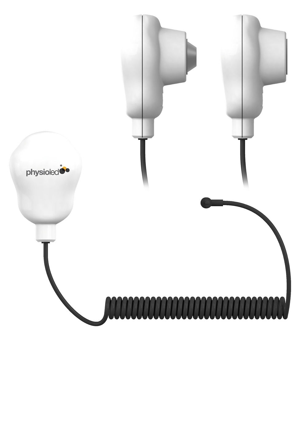
The Sensor Medica Moover Gait™ is a professional wearable wireless network of inertial measurement units (IMUs) for complete three-dimensional gait analysis and range of motion (ROM) assessment
Moover Gait is Wearable — wireless, non-invasive, without external infrastructures. It is Accurate — validated with gold-standard optoelectronic systems. It is Innovative — with real-time 3D viewing and video synchronization.
Analysis Outputs — What Moover Gait Measures
Raw Data Visualization Visualization of the Timeline of raw data, joint angles, video and 3D animation. The raw data timeline displays the continuous sensor output — acceleration, angular velocity, and orientation — alongside computed joint angle curves and the synchronized 3D body animation and video, enabling the clinician to review the complete acquisition in detail before report generation. Medicalexpo
Spatiotemporal Gait Parameters Moover Gait computes the core space-time measurements that characterize gait quality and symmetry:
- Symmetry indices of duration and length of the half step and step — quantifying left/right step length and timing asymmetry Medicalexpo
- Symmetry indices of support times during the gait phases — stance phase, swing phase, and double support symmetry between limbs Medicalexpo
- Symmetry indices of the impact forces of the leg and foot — loading asymmetry at initial contact Medicalexpo
- Spatio-temporal indices of the steps of the walk — speed, times and lengths — walking speed, step length, stride length, step time, and stride time Medicalexpo
- Step cadence — steps per minute Medicalexpo
These spatiotemporal symmetry indices are the clinical core of gait rehabilitation monitoring — directly quantifying the asymmetries that visual observation can only estimate, and tracking their reduction over the course of a rehabilitation programme.
3D Joint Kinematics — Pelvis, Hip, Knee & Ankle 3D joint angles of the pelvis, hip, knee and ankle are computed in all three planes — sagittal, frontal, and transverse — providing the complete lower limb kinematic profile throughout the gait cycle. Joint angle curves are displayed for each phase of the gait cycle and compared against normative ranges, enabling the clinician to identify: Medicalexpo
- Hip flexion/extension deficits and asymmetries
- Excessive or insufficient hip abduction/adduction
- Hip rotation abnormalities in transverse plane
- Knee flexion during loading response (shock absorption)
- Knee hyperextension in mid and terminal stance
- Contralateral pelvic drop (Trendelenburg sign)
- Pelvic rotation and obliquity abnormalities
- Ankle dorsiflexion and plantarflexion through stance and swing
- Ankle frontal plane deviation (valgus/varus)
Angular Symmetry Indices Angular symmetry indices are computed from the 3D joint angle data — providing a single quantitative metric for left/right kinematic symmetry at each joint and in each plane. These indices are particularly valuable for post-surgical rehabilitation (e.g., total knee arthroplasty, ACL reconstruction) and neurological rehabilitation (stroke hemiplegia) where restoration of joint angle symmetry is a primary treatment outcome. Medicalexpo
Automatic Gait Analysis Report (PDF) The gait analysis report in PDF is automatically generated by the software. It includes spatiotemporal parameters, symmetry indices, joint angle curves in each plane, angular symmetry indices, as well as detailed information on the mobility of each joint and its Normal Range. The automated PDF report is generated immediately following assessment — requiring no manual data compilation, no graph construction, and no report-writing time. The report includes normative range comparison for each joint parameter, enabling any clinician to identify clinically significant deviations without specialist gait analysis training. Medicalexpo
Automatic ROM Analysis Report (PDF) The ROM analysis report in PDF is automatically generated by the software. It includes angles, angular velocity, angular acceleration, and graphs of the selected joints for a kinematic study of the analyzed movement. The ROM report extends Moover Gait's capability beyond gait analysis into general joint range of motion assessment — providing objective, repeatable kinematic ROM data for clinical physiotherapy assessment, pre/post-treatment comparison, and rehabilitation outcome monitoring. Medicalexpo
3D Data Export in BVH Format 3D export of data in BVH (Biovision Hierarchy format) — the standard skeletal animation data format compatible with motion analysis research software, biomechanical modelling tools, and 3D animation applications. BVH export enables Moover Gait data to be used in research workflows, multi-system clinical analyses, and specialist biomechanical applications beyond the freeStep environment.
Quantity
Moover Gait vs. Optoelectronic Motion Capture —
| Parameter | Moover Gait™ (IMU) | Optoelectronic Motion Capture |
|---|---|---|
| Setup time | Minutes | 30–60 minutes |
| Infrastructure required | None — wireless, wearable | Camera arrays, lab space, force plates |
| Portability | Full — any environment | Fixed laboratory |
| Outdoor / community use | Yes | Not practical |
| Validated accuracy | Validated vs optoelectronic | Gold standard reference |
| Real-time 3D visualization | Yes | Yes |
| 3D joint kinematics | Pelvis, hip, knee, ankle | Full body including upper limb |
| Force/kinetics | Not included | With force plates |
| Cost | Significantly lower | Very high |
RELATED PRODUCTS